Defibrillation
Defibrillation is the definitive treatment for the life-threatening cardiac arrhythmias, ventricular fibrillation and pulseless ventricular tachycardia. Defibrillation consists of delivering a therapeutic dose of electrical energy to the affected heart with a device called a defibrillator. This depolarizes a critical mass of the heart muscle, terminates the arrhythmia, and allows normal sinus rhythm to be reestablished by the body's natural pacemaker, in the sinoatrial node of the heart. Defibrillators can be external, transvenous, or implanted, depending on the type of device used or needed. Some external units, known as automated external defibrillators (AEDs), automate the diagnosis of treatable rhythms, meaning that lay responders or bystanders are able to use them successfully with little, or in some cases no training at all.
History
Defibrillation was first demonstrated in 1899 by Jean Louis Prevost and Frederic Batelli, two physiologists from University of Geneva, Switzerland. They discovered that small electric shocks could induce ventricular fibrillation in dogs, and that larger charges would reverse the condition.
In 1933 a Dr Albert Hyman a heart specialist at the Beth Davis Hospital of New York city and a C. Henry Hyman, an electrical engineer, looking for an alternative to injecting powerful drugs directly into the heart, came up with an invention that used an electrical shock in place of drug injection. This invention was called the Hyman Otor where a hollow needle is used to pass an insulated wire to the heart area to deliver the electrical shock. The hollow steel needle being one end of the circuit and the insulated wire the other end. Whether the Hyman Otor was a success is unknown. [1]
The first use on a human was in 1947 by Claude Beck,[2] professor of surgery at Case Western Reserve University. Beck's theory was that ventricular fibrillation often occurred in hearts which were fundamentally healthy, in his terms "Hearts that are too good to die", and that there must be a way of saving them. Beck first used the technique successfully on a 14 year old boy who was being operated on for a congenital chest defect. The boy's chest was surgically opened, and manual cardiac massage was undertaken for 45 minutes until the arrival of the defibrillator. Beck used internal paddles on either side of the heart, along with procainamide, an antiarrhythmic drug, and achieved return of normal sinus rhythm.
These early defibrillators used the alternating current from a power socket, transformed from the 110-240 volts available in the line, up to between 300 and 1000 volts, to the exposed heart by way of 'paddle' type electrodes. The technique was often ineffective in reverting VF while morphological studies showed damage to the cells of the heart muscle post mortem. The nature of the AC machine with a large transformer also made these units very hard to transport, and they tended to be large units on wheels.
Closed-chest method
Until the early 1950s, defibrillation of the heart was possible only when the chest cavity was open during surgery. The technique used an alternating current from a 300 or greater volt source delivered to the sides of the exposed heart by 'paddle' electrodes where each electrode was a flat or slightly concave metal plate of about 40 mm diameter. The closed-chest defibrillator device which applied an alternating current of greater than 1000 volts, conducted by means of externally applied electrodes through the chest cage to the heart, was pioneered by Dr V. Eskin with assistance by A. Klimov in Frunze, USSR (today known as Bishkek, Kyrgyzstan) in mid 1950s.
Move to direct current
In 1959 Bernard Lown commenced research into an alternative technique which involved charging of a bank of capacitors to approximately 1000 volts with an energy content of 100-200 joules then delivering the charge through an inductance such as to produce a heavily damped sinusoidal wave of finite duration (~5 milliseconds) to the heart by way of 'paddle' electrodes. The work of Lown was taken to clinical application by engineer Barouh Berkovits with his "cardioverter".
The Lown waveform, as it was known, was the standard for defibrillation until the late 1980s when numerous studies showed that a biphasic truncated waveform (BTE) was equally efficacious while requiring the delivery of lower levels of energy to produce defibrillation. A side effect was a significant reduction in weight of the machine. The BTE waveform, combined with automatic measurement of transthoracic impedance is the basis for modern defibrillators.
Portable units become available
A major breakthrough was the introduction of portable defibrillators used out of the hospital. This was pioneered in the early 1960s by Prof. Frank Pantridge in Belfast. Today portable defibrillators are among the many very important tools carried by ambulances. They are the only proven way to resuscitate a person who has had a cardiac arrest unwitnessed by EMS who is still in persistent ventricular fibrillation or ventricular tachycardia at the arrival of pre-hospital providers.
Gradual improvements in the design of defibrillators, partly based on the work developing implanted versions (see below), have led to the availability of Automated External Defibrillators. These devices can analyse the heart rhythm by themselves, diagnose the shockable rhythms, and charge to treat. This means that no clinical skill is required in their use, allowing lay people to respond to emergencies effectively.
Implantable devices
A further development in defibrillation came with the invention of the implantable device, known as an implantable cardioverter-defibrillator (or ICD). This was pioneered at Sinai Hospital in Baltimore by a team that included Stephen Heilman, Alois Langer, Jack Lattuca, Morton Mower, Michel Mirowski, and Mir Imran, with the help of industrial collaborator Intec Systems of Pittsburgh.[5] Mirowski teamed up with Mower and Staewen, and together they commenced their research in 1969 but it was 11 years before they treated their first patient. Similar developmental work was carried out by Schuder and colleagues at the University of Missouri.
The work was commenced, despite doubts amongst leading experts in the field of arrhythmias and sudden death. There was doubt that their ideas would ever become a clinical reality. In 1962 Bernard Lown introduced the external DC defibrillator. This device applied a direct current from a discharging capacitor through the chest wall into the heart to stop heart fibrillation.[6] In 1972, Lown stated in the journal Circulation - "The very rare patient who has frequent bouts of ventricular fibrillation is best treated in a coronary care unit and is better served by an effective antiarrhythmic program or surgical correction of inadequate coronary blood flow or ventricular malfunction. In fact, the implanted defibrillator system represents an imperfect solution in search of a plausible and practical application."[7]
The problems to be overcome were the design of a system which would allow detection of ventricular fibrillation or ventricular tachycardia. Despite the lack of financial backing and grants, they persisted and the first device was implanted in February 1980 at Johns Hopkins Hospital by Dr. Levi Watkins, Jr. Modern ICDs do not require a thoracotomy and possess pacing, cardioversion, and defibrillation capabilities.
The invention of implantable units is invaluable to some regular sufferers of heart problems, although they are generally only given to those people who have already had a cardiac episode.
Manual external defibrillator
The units are used in conjunction with (or more often have inbuilt) electrocardiogram readers, which the healthcare provider uses to diagnose a cardiac condition (most often fibrillation or tachycardia although there are some other rhythms which can be treated by different shocks). The healthcare provider will then decide what charge (in joules) to use, based on proven guidelines and experience, and will deliver the shock through paddles or pads on the patient's chest. As they require detailed medical knowledge, these units are generally only found in hospitals and on some ambulances. For instance, every NHS ambulance in the United Kingdom is equipped with a manual defibrillator for use by the attending paramedics and technicians. In the United States, many advanced EMTs and all paramedics are trained to recognize lethal arrhythmias and deliver appropriate electrical therapy with a manual defibrillator when appropriate.
Automated external defibrillator (AED)
These simple-to-use units are based on computer technology which is designed to analyze the heart rhythm itself, and then advise the user whether a shock is required. They are designed to be used by lay persons, who require little training to operate them correctly. They are usually limited in their interventions to delivering high joule shocks for VF (ventricular fibrillation) and VT (ventricular tachycardia) rhythms, making them generally of limited use to health professionals, who could diagnose and treat a wider range of problems with a manual or semi-automatic unit.
The automatic units also take time (generally 10–20 seconds) to diagnose the rhythm, where a professional could diagnose and treat the condition far more quickly with a manual unit.[8] These time intervals for analysis, which require stopping chest compressions, have been shown in a number of studies to have a significant negative effect on shock success.[9] This effect led to the recent change in the AHA defibrillation guideline (calling for two minutes of CPR after each shock without analyzing the cardiac rhythm) and some bodies recommend that AEDs should not be used when manual defibrillators and trained operators are available.[8]
Automated external defibrillators are generally either held by trained personnel who will attend incidents, or are public access units which can be found in places including corporate and government offices, shopping centres, airports, restaurants, casinos, hotels, sports stadiums, schools and universities, community centers, fitness centers and health clubs.
The locating of a public access AED should take in to account where large groups of people gather, and the risk category associated with these people, to ascertain whether the risk of a sudden cardiac arrest incident is high. For example, a center for teenage children is a particularly low risk category (as children very rarely enter heart rhythms such as VF (Ventricular Fibrillation) or VT (Ventricular Tachycardia), being generally young and fit, and the most common causes of pediatric cardiac arrest are respiratory arrest and trauma - where the heart is more likely to enter asystole or PEA, (where an AED is of no use). On the other hand, a large office building with a high ratio of males over 50 is a very high risk environment.
In many areas, emergency services vehicles are likely to carry AEDs. EMT-Basics in most areas are not trained in manual defibrillation, and often carry an AED instead. Some ambulances carry an AED in addition to a manual unit. In addition, some police or fire service vehicles carry an AED for first responder use. Some areas have dedicated community first responders, who are volunteers tasked with keeping an AED and taking it to any victims in their area. It is also increasingly common to find AEDs on transport such as commercial airlines and cruise ships. The presence of an AED can be a particularly decisive factor in cardiac patient survival in these scenarios, as professional medical assistance may be hours away.
In order to make them highly visible, public access AEDs often are brightly coloured, and are mounted in protective cases near the entrance of a building. When these protective cases are opened, and the defibrillator removed, some will sound a buzzer to alert nearby staff to their removal but do not necessarily summon emergency services. All trained AED operators should also know to phone for an ambulance when sending for or using an AED, as the patient will be unconscious, which always requires ambulance attendance.
Implantable cardioverter-defibrillator (ICD)
Main article: Implantable cardioverter-defibrillator
Also known as automatic internal cardiac defibrillator (AICD). These devices are implants, similar to pacemakers (and many can also perform the pacemaking function). They constantly monitor the patient's heart rhythm, and automatically administer shocks for various life threatening arrhythmias, according to the device's programming. Many modern devices can distinguish between ventricular fibrillation, ventricular tachycardia, and more benign arrhythmias like supraventricular tachycardia and atrial fibrillation. Some devices may attempt overdrive pacing prior to synchronised cardioversion. When the life threatening arrhythmia is ventricular fibrillation, the device is programmed to proceed immediately to an unsynchronized shock.
There are cases where the patient's ICD may fire constantly or inappropriately. This is considered a medical emergency, as it depletes the device's battery life, causes significant discomfort and anxiety to the patient, and in some cases may actually trigger life threatening arrhythmias. Some emergency medical services personnel are now equipped with a ring magnet to place over the device, which effectively disables the shock function of the device while still allowing the pacemaker to function (if the device is so equipped). If the device is shocking frequently, but appropriately, EMS personnel may administer sedation.
Wearable cardiac defibrillator
A development of the AICD is a portable external defibrillator that is worn like a vest.[10] The unit monitors the patient 24 hours a day and will automatically deliver a biphasic shock if needed. This device is mainly indicated in patients awaiting an implantable defibrillator. Currently only one company manufactures these and they are of limited availability.
Interface with the patient
The most well-known type of electrode (widely depicted in films and television) is the traditional metal paddle with an insulated (usually plastic) handle. This type must be held in place on the patient's skin while a shock or a series of shocks is delivered. Before the paddle is used, a gel must be applied to the patient's skin, in order to ensure a good connection and to minimize electrical resistance, also called chest impedance (despite the DC discharge). These are generally only found on the manual external units.
Newer types of resuscitation electrodes are designed as an adhesive pad. These are peeled off their backing and applied to the patient's chest when deemed necessary, much the same as any other sticker. These electrodes are then connected to a defibrillator. If defibrillation is required, the machine is charged, and the shock is delivered, without any need to apply any gel or to retrieve and place any paddles. These adhesive pads are found on most automated and semi-automated units, and are gradually replacing paddles entirely in non-hospital settings.
Both solid- and wet-gel adhesive electrodes are available. Solid-gel electrodes are more convenient, because there is no need to clean the patient's skin after removing the electrodes. However, the use of solid-gel electrodes presents a higher risk of burns during defibrillation, since wet-gel electrodes more evenly conduct electricity into the body.
Some adhesive electrodes are designed to be used not only for defibrillation, but also for transcutaneous pacing and synchronized electrical cardioversion.
In a hospital setting, paddles are generally preferred to pads, due to the inherent speed with which they can be placed and used. This is critical during cardiac arrest, as each second of nonperfusion means tissue loss. However, in cases in which cardiac arrest is suspected, patches placed prophalactically are superior,as they provide appropriate EKG tracing without the artifact visible from human interference with the paddles. Adhesive electrodes are also inherently safer than the paddles for the operator of the defibrillator to use, as they minimize the risk of the operator coming into physical (and thus electrical) contact with the patient as the shock is delivered, by allowing the operator to stand several feet away. Adhesive patches also require no force to remain in place and deliver the shock appropriately, whereas paddles require approximately 25 lbs of force to be applied while the shock is delivered.
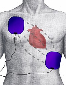
Placement
Anterio-apical placement of external defibrillator electrodes (When defibrillation is unsuccessful, anterior-posterior placement is also sometimes attempted)
Resuscitation electrodes are placed according to one of two schemes. The anterior-posterior scheme (conf. image) is the preferred scheme for long-term electrode placement. One electrode is placed over the left precordium (the lower part of the chest, in front of the heart). The other electrode is placed on the back, behind the heart in the region between the scapula. This placement is preferred because it is best for non-invasive pacing.
The anterior-apex scheme can be used when the anterior-posterior scheme is inconvenient or unnecessary. In this scheme, the anterior electrode is placed on the right, below the clavicle. The apex electrode is applied to the left side of the patient, just below and to the left of the pectoral muscle. This scheme works well for defibrillation and cardioversion, as well as for monitoring an ECG.
Popular culture references
As devices that can quickly produce dramatic improvements in patient health, defibrillators are often depicted in movies, television, video games and other fictional media. Their function, however, is often exaggerated, with the defibrillator inducing a sudden, violent jerk or convulsion by the patient; in reality, although the muscles may contract, such dramatic patient presentation is rare. Similarly, medical providers are often depicted defibrillating patients with a "flat-line" ECG rhythm (also known as asystole); this is not done in real life. Only the cardiac arrest rhythms ventricular fibrillation and pulseless ventricular tachycardia are normally defibrillated. (There are also several heart rhythms that can be "shocked" when the patient is not in cardiac arrest, such as supraventricular tachycardia and ventricular tachycardia that produces a pulse; this procedure is known as cardioversion, not defibrillation.)
In Australia up until the 1990s it was quite rare for ambulances to carry defibrillators. This changed in 1990 after Australian media mogul Kerry Packer had a heart attack and, purely by chance, the ambulance that responded to the call carried a defibrillator. After recovering, Kerry Packer donated a large sum to the Ambulance Service of New South Wales in order that all ambulances in New South Wales should be fitted with a personal defibrillator, which is why defibrillators in Australia are colloquially called "Packer Whackers".
No comments:
Post a Comment